,
Newly Discovered Shock
Absorber in the Equine Foot (7-24-07)
Pete Ramey
Important note: These are just
preliminary observations. They are my interpretation after
several conversations about it with Dr. Bowker. The completed
research project is coming eventually, but people who went to
his last clinic are buzzing about it, so I thought I'd try to
clear it up.
Robert Bowker VMD, PhD has been teaching
for many years that the blood flow in the equine foot acts as a
hydraulic shock absorber. Most of his focus
has been on the back half of the foot, but more recently he's
paying more attention to energy dissipating features in the
front half of the foot as well.
Recent data shows that peripheral loading
of the foot (unloading the sole) reduces hoof perfusion by
almost 50%.... Immediately. This does not necessarily cause
tissue death, because the sole's corium is filled with a huge
number of micro-vessels – way more than are needed for healthy
tissue life. Bowker feels these 'extra' blood vessels are for
hydraulic energy dissipation, but more recently he's discovered
that the entire structure of the sole's corium is a mixture of
venous microvasculature surrounded by proteoglycans – an
extremely elastic structure (along with a "honeycomb" framework
of keratinized sole). This type of structure is known to have
"use it or lose it" tendencies. The more it is used the better
it develops – the more elastic it becomes.
Bowker has noticed that unhealthy or
under-developed equine feet have a thin solar corium that is
fairly uniform all the way across (1-3 mm), but healthy,
well-developed feet have a much thicker corium in the outer
periphery. This thicker corium may be 3-5 mm thick (or more) in
the healthiest hooves.
Aside from a tremendous "Gel Pad" shock
absorber, this thicker corium also allows for a great deal of
expansion room of the front half of the
foot. This is very significant, as many people still think the
expansion only happens in the back half of the foot; where the
foundation for the hoof capsule is cartilage instead of bone.
The pictures below are 10mm thick slices
taken 12mm behind the apex of the frog. Notice as I apply hard
pressure with my hand, the solar corium flattens, the frog moves
to the ground and the walls spread dramatically. The force
required to do this to the thin section is basically "as hard as
I can push." As this is studied more, we'll elaborate, but I
thought you'd like to hear about it now. Pete
The walls can spread
significantly as pressure is applied to P3 and the sole flattens. The
thicker corium at the distal border of P3 is compressed, pushing blood to
the back of the foot through an energy dampening network of micro-vessels.
Then when the load is released, the elastic nature of the sole's corium and
spring tension in the hoof capsule snaps it all back into place for
the next stride. (These pictures are the exact same size, of the same slice,
and taken from the exact same range, 2 second time lapse.)
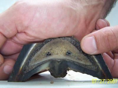
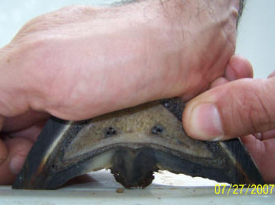
At Rest Applying Hard Pressure
Also note that this pressure
does not create a separational force on the laminae – they actually
compress! If the wall was not allowed to expand, the same downward force
would stretch the laminae.
The thin corium at the center
of P3 seems to thicken with weight bearing, as the corium at the outer
periphery is compressed.