
Reading Sole Thickness
Q: Hello Pete,
Is there an accurate
way to estimate the thickness of a horse’s sole?
A: Very good question! 20 years ago, I was
actually trained to trim the sole until tiny dots of blood appeared (and I still
see people doing this today). I soon realized that when blood appears, you have
already trimmed at least a half-inch too far—removing all of the horse’s natural
protection of the sensitive corium of the foot. When trimming a horse’s foot, we
should
always be careful to leave this
1/2- to 3/4-inch-thick layer of protective sole, but we must also remove any
excess to allow the foot to be as compact as possible to maintain agility. This
means that estimating sole thickness is crucial to competent trimming and
shoeing.
Important Note: The
very best you can do is no more, and no less than an estimation! When in doubt,
or if the situation is critical, always get radiographs to be sure. That said,
even when the highest-quality radiographs are available, it is really only
possible to measure sole thickness at a small part of the sole that is in
profile in the image. Additionally, the radiograph shows the sole thickness only
during the moment in time (in the past) that it was taken—so the estimation of
sole thickness is still important.
The first and most important step in estimating sole
thickness is developing an understanding of the shape of the internal
structures. The “foundation” of the
front-half of the foot is bone—the
foundation of the back-half is flexible cartilage. Together, they form the
shape of a miniature hoof with a concaved sole. Covering this bone and cartilage
“foundation” is a 3/8th-inch-thick sock-like layer of live, vascular,
nerve-filled tissue (see figure 1). This layer—the corium—must always be
well-protected.
Cadaver specimen.
This is how the horses’ foot looks with the “skin” (hoof wall, sole, bars and
frog) removed. All of this highly-sensitive tissue must be covered by at least a
1/2-inch-thick protective layer for the horse to be safe and sound. If the
slightest bit of blood is drawn during a trim, a significant error in judgment
was made—not a slight slip-up. Photo reprinted from the book Care and
Rehabilitation of the Equine Foot, P. Ramey.
The shape of these internal structures can vary. Some
horses may have slightly more or less concavity of the solar surface. Other
horses—particularly past founder cases and horses with upright or “club”
feet—may have a remodeled coffin bone, altering this shape at the toe area. This
is why radiographs remain important, even for highly-skilled estimators of sole
thickness. But most horses will have a general shape and form very similar to
the Figure 1. Looking at this picture, take special note of the area between the
frog corium and the sole corium. This area forms the collateral grooves—the
“seam” between the frog and sole that you typically clean out with a hoof pick.
The collateral grooves are important for sole estimation because in a normal
hoof, the sole material in the bottom of the groove rarely varies in thickness.
The very deepest part of this groove tends to be 3/8th-inch away from
the corium (live tissue) whether the rest of the sole is too thick or too thin.
This means that from one end to the other, the collateral grooves along the frog
are quite accurate for locating the
sensitive tissue. Exceptions: When
subsolar abscessing has been present, the collateral grooves may be farther from
the corium. Fungal and/or bacterial infections may erode them closer or all the
way into the corium. Always be prepared to fall back on radiographs when
conditions of the foot are abnormal.
So putting it all together so far, if 1) you know how the
internal structures are shaped, and 2) you know where they are within the foot,
it becomes pretty easy to estimate the thickness of the sole in any area of the
foot.
To use this insight in the field, be sure that there is
always enough sole material (in the outer perimeter adjacent to the white line)
to lift the collateral grooves at least ½-inch (or more) off the ground. This
can be measured by picking up the foot and laying a rasp across the foot—in any
direction, depending on which part of the sole you are evaluating. Measure from
the rasp to the bottom of the collateral groove with a thin ruler (see Figure
2). The groove should generally be left deeper in the back of the foot (heel
height is a separate and complex subject) and the sole at the toe should never
be trimmed so that the collateral groove at the apex of the frog is less than
½-inch off the ground—5/8ths-inch is even better, plus any additional lift
provided by hoof wall extending past the sole.
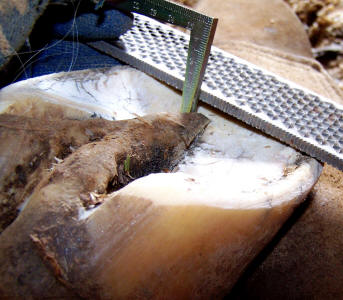
Proper placement of rasp and ruler to estimate the thickness of sole in the
areas where the rasp has been placed. If the measurement to the bottom of the
groove is 1/2- to 3/4-inch, there is probably a correct amount of sole covering
the internal structures. Photo reprinted from the book Care and
Rehabilitation of the Equine Foot, P. Ramey.
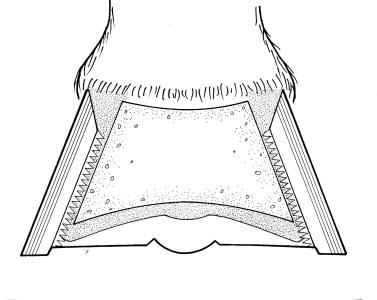
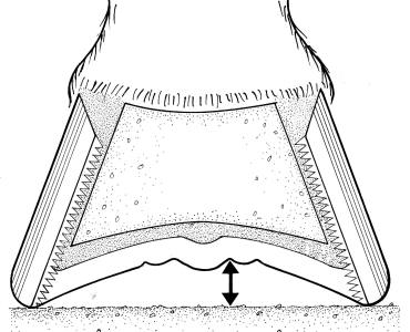
These drawings of the hoof, sectioned crossways just behind the apex of the frog
(as if the toe was chopped off and you were looking at it from the front). Shown
are the coffin bone, surrounding live corium, hoof wall and protective sole and
frog material. Notice in the left photo, the collateral groove would be very
close to the ground because the sole adjacent to the hoof wall is very thin. In
the right photo, the sole is thicker, lifting the collateral groove higher off
the ground. Drawings by Karen Sullivan, reprinted from the book Care and
Rehabilitation of the Equine Foot, P. Ramey.
Once you can visualize sole thickness using the collateral
grooves, the next step will make you more accurate and somewhat able to predict
bone remodeling and/or thin areas of the sole. The horse’s sole
tends to callus into a fairly uniform thickness covering the live tissue
in seen figure 1. Therefore, the sole you see when you pick up the horse’s foot
should be concaved into a bowl-shape with no flattened areas adjacent to the
white line. If you find that a horse’s sole is flat (instead of concaved)
immediately adjacent to the white line, you have either 1) a thinner area of
sole in the flattened region, or 2) a remodeled coffin bone in that region.
Generally, a radiograph is required to tell the difference between the
two, but either way, the area should probably not be thinned more than it
already is.
When these two methods (collateral groove height and
reading the shape of the sole) are combined, they give a quite accurate
estimation of sole thickness all over the bottom of the foot—you can see for
yourself if someone is over-thinning your horse’s sole or trimming areas of the
foot that are already too short. Learning to read the sole will also help you
keep your horses out of trouble by allowing you to notice if they excessively
wear a foot or develop a reduced capacity for sole growth. Protect these feet
with less-abrasive terrain and/or some protective device
before their sensitive tissues are
damaged.